Spondylolisthesis and spinal stenosis
Spondylolisthesis?

Degenerative change in the vertebrae, severe disc damage, or a congenital defect in the spinal arch can produce a condition in which the upper vertebra slips forward relative to the one below.
It occurs mainly in the lumbar spine, and when it compresses surrounding nerves and structures, pain and neurological symptoms can result.
Those who repeatedly adopt postures and habits that are hard on the lower back should take care, as symptoms can appear more readily than in others.
What is spinal stenosis, and how does it differ from lumbar disc disease?
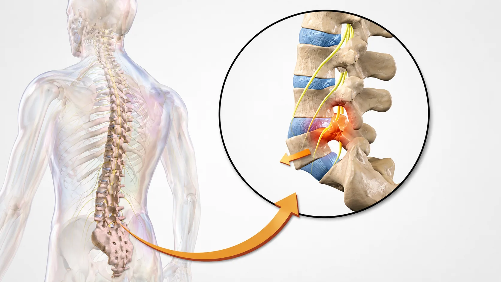
This is a condition in which the spinal canal — the space the nerves pass through — narrows and chronically compresses them. Lumbar disc disease and spinal stenosis resemble each other in that compression of the lumbar nerves running from the back into the leg causes numbness and impairs walking.
In lumbar disc disease, however, the usual finding is radiating pain caused by damaged or herniated disc material — the annulus or nucleus — pressing on a nerve. In chronic spinal stenosis, by contrast, hard tissue such as thickened ligament and bone spurs also presses on the nerve in addition to the damaged disc.
Lumbar disc symptoms usually begin acutely, whereas spinal stenosis more often develops gradually over a long period. A further characteristic symptom of spinal stenosis is neurogenic claudication. This distinguishes it from ordinary lumbar disc disease: the patient is comfortable while seated, but after walking only a short distance the legs become numb and painful, requiring a rest before walking again. Because it resembles peripheral vascular disease, testing is needed to tell them apart.
Check the main symptoms of spinal stenosis
| Worse on walking · eases with rest | Legs become weak or painful after walking or standing for a long time, and the symptoms ease with rest |
|---|---|
| Eases on bending forward | When bending forward reduces the pain |
| Persistent low back pain | Persistent low back pain that worsens on standing or walking for long periods |
| Numbness · Altered Sensation | Altered sensation such as numbness or dullness in the leg or buttock |
From diagnosis to treatment — here is how it works
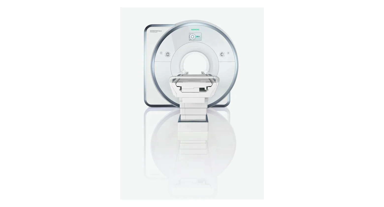
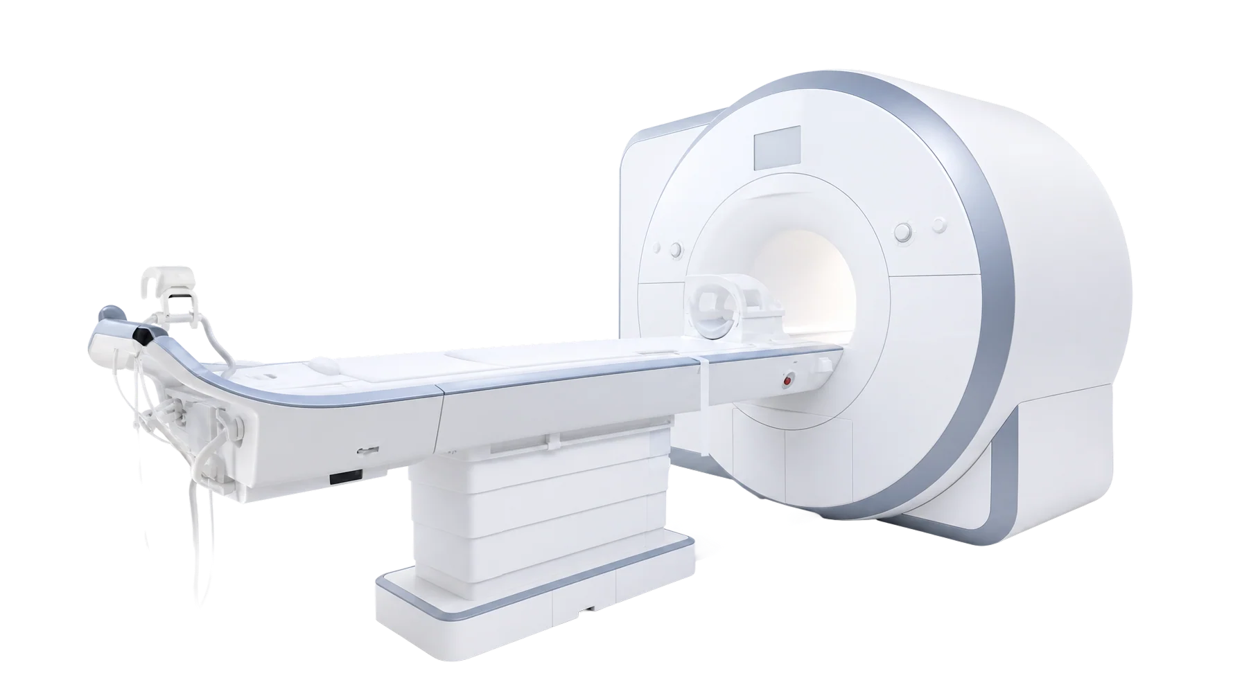
High-resolution cervical or lumbar MRI identifies which disc level is damaged and shows the underlying spinal structure. Nerve conduction and EMG studies pinpoint exactly which nerve is compressed, and with that information most patients can expect improvement with non-surgical treatment.
At our clinic we select only what is needed from epidural nerve block, selective nerve root injection, intradiscal anti-inflammatory treatment, medication, and physical therapy and proceed step by step.
History & Physical Examination
In most cases, history-taking together with physical examination is sufficient.
MRI if needed
MRI is added when symptoms are severe or a precise diagnosis is required.
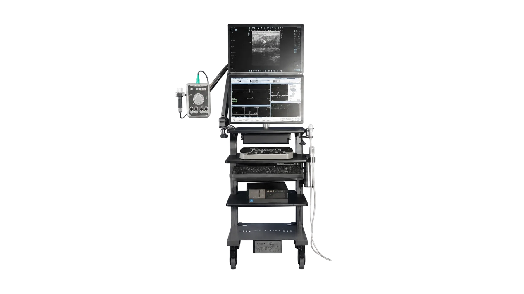
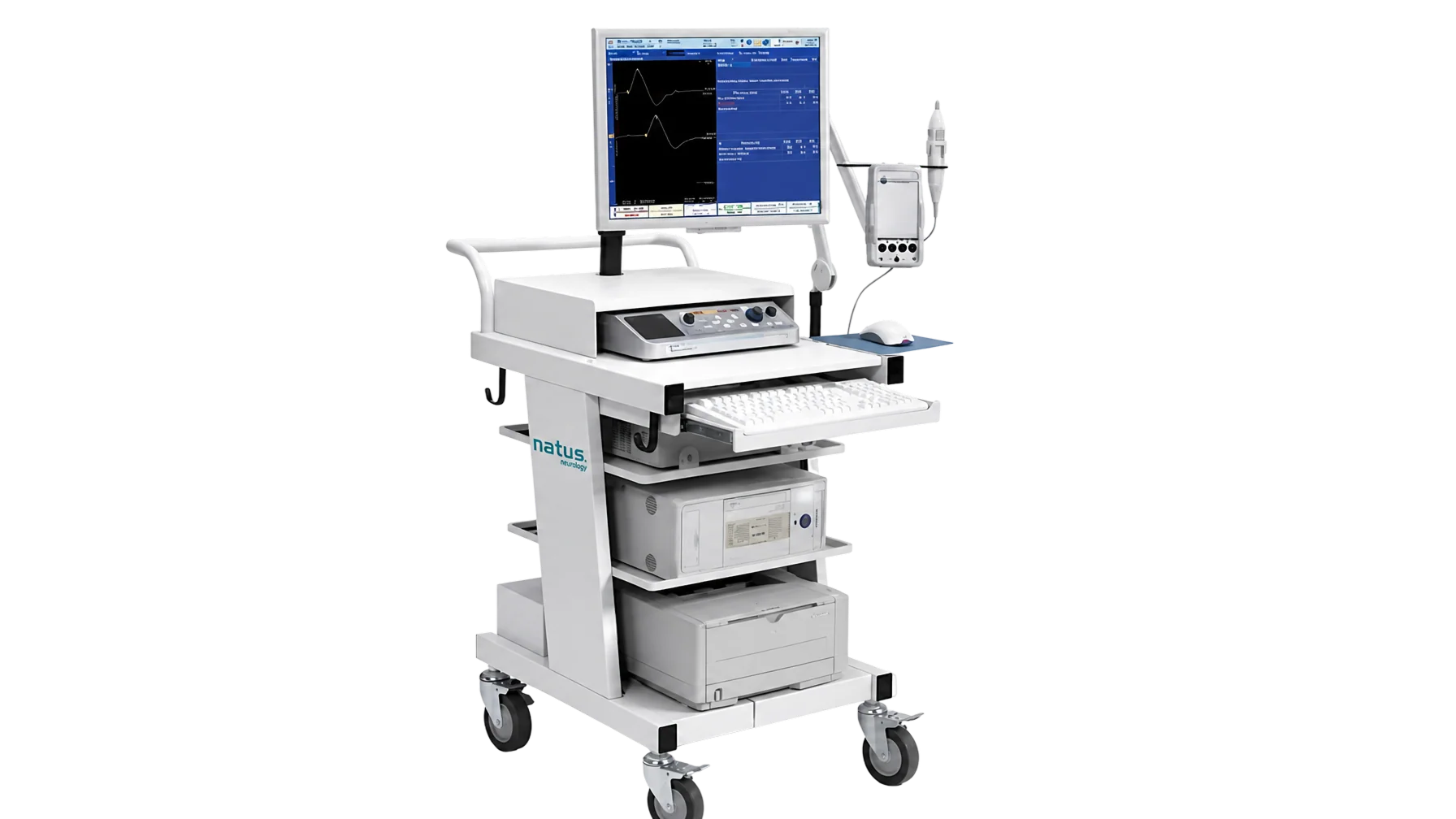
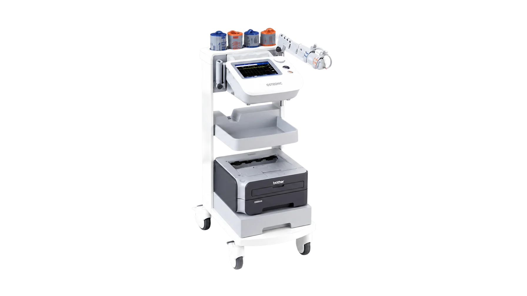
Nerve Conduction & EMG
May be needed to rule out other conditions or to determine the exact level and severity of nerve root compression.
Medication & Rehabilitation Therapy
Nerve Block · Anti-inflammatory Treatment
When pain is severe enough to hinder rehabilitation and recovery, nerve block and peri-discal anti-inflammatory treatment may be considered.
※ This is general medical information provided for understanding only. Actual diagnosis and treatment are determined by a specialist after examination, based on the individual patient's condition.