Spinal nerve treatment (epidural and nerve root injection)

Spinal Nerve Block
Spinal Nerve Block
Spinal Nerve Block

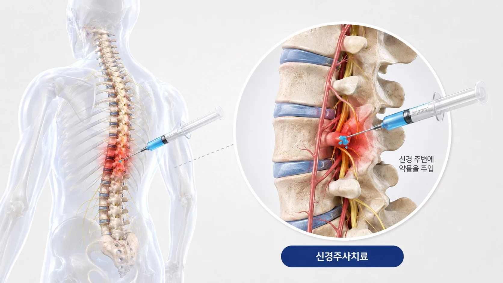
Severe pain often results when nerves running into the limbs are compressed by disc damage or foraminal stenosis, or by inflammation and swelling.
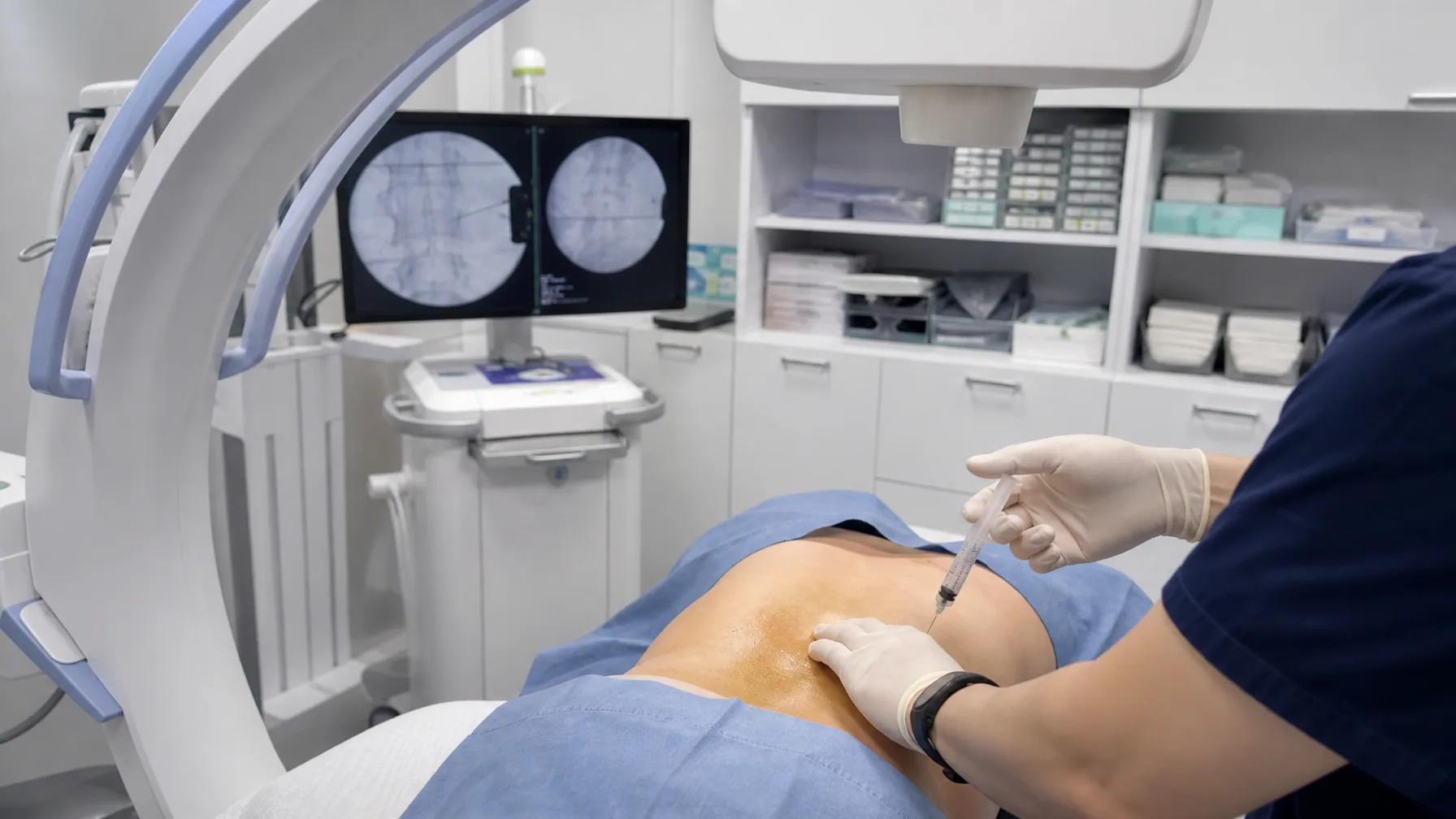
In such cases Medication is delivered directly to the compressed nerve root or the epidural space under real-time imagingreduces inflammation and swelling and blocks the pathways transmitting harmful pain.
The procedure is brief, requires no anesthesia, and no hospital stay.

Spinal nerve treatment or When selective nerve root block is required

Severe painWhen severe neck or back pain limits daily life
Hinders rehabilitationWhen severe pain is judged to be hindering rehabilitation and recovery
Radiating painNeck or back pain together with severe numbness radiating into the buttock, leg, or arm
Positional painAching and worsening pain around both shoulders or hips after sitting in a poor posture for long periods or after walking
When surgery is difficultSevere spinal and radiating limb pain where surgery is not possible or not wanted

Precautions before and after the procedure

1. A small dose of steroid is used only when inflammatory pain is severe. Steroids must be used when genuinely indicated, but repeated high doses carry side effects, so discuss this with your physician.
2. The injection may temporarily paralyze a motor nerve. This generally resolves within a week.
3. When local anesthetic is used in large amounts, some may be absorbed into the bloodstream and cause systemic symptoms immediately after injection, though dangerous reactions are generally not seen.

※ This is general medical information provided for understanding only. Whether and how to treat is determined by a specialist after examination, based on the individual patient's condition.
Advanced Equipment

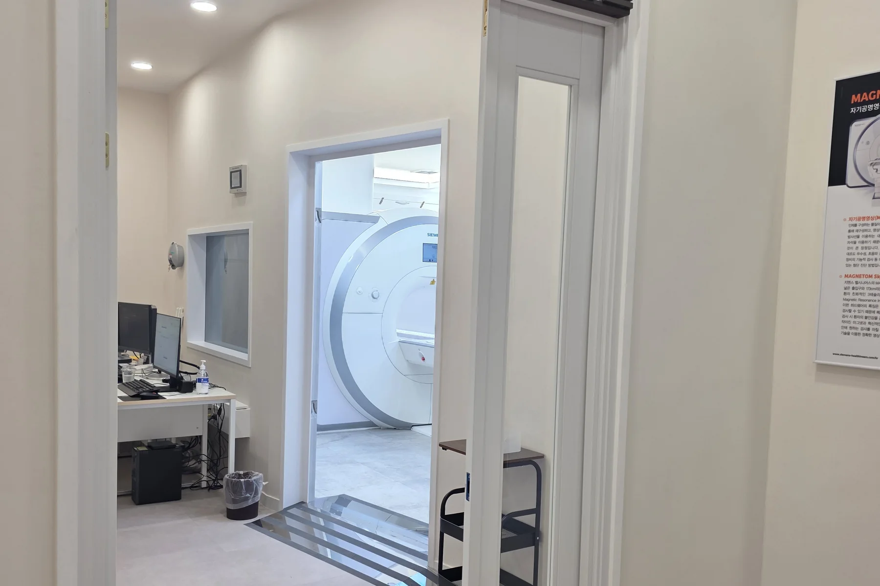
  • SIEMENS3T AI MRIWe add AI imaging (Deep Resolve) to a proven Siemens 3T MRI to examine neurological conditions faster and more clearly.
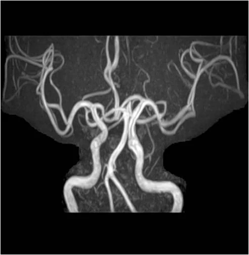
  • MRACerebrovascular and carotid MRAIt accurately evaluates the degree of atherosclerosis, stenosis, structural abnormality, and the presence of aneurysm in the vessels of the brain and neck.
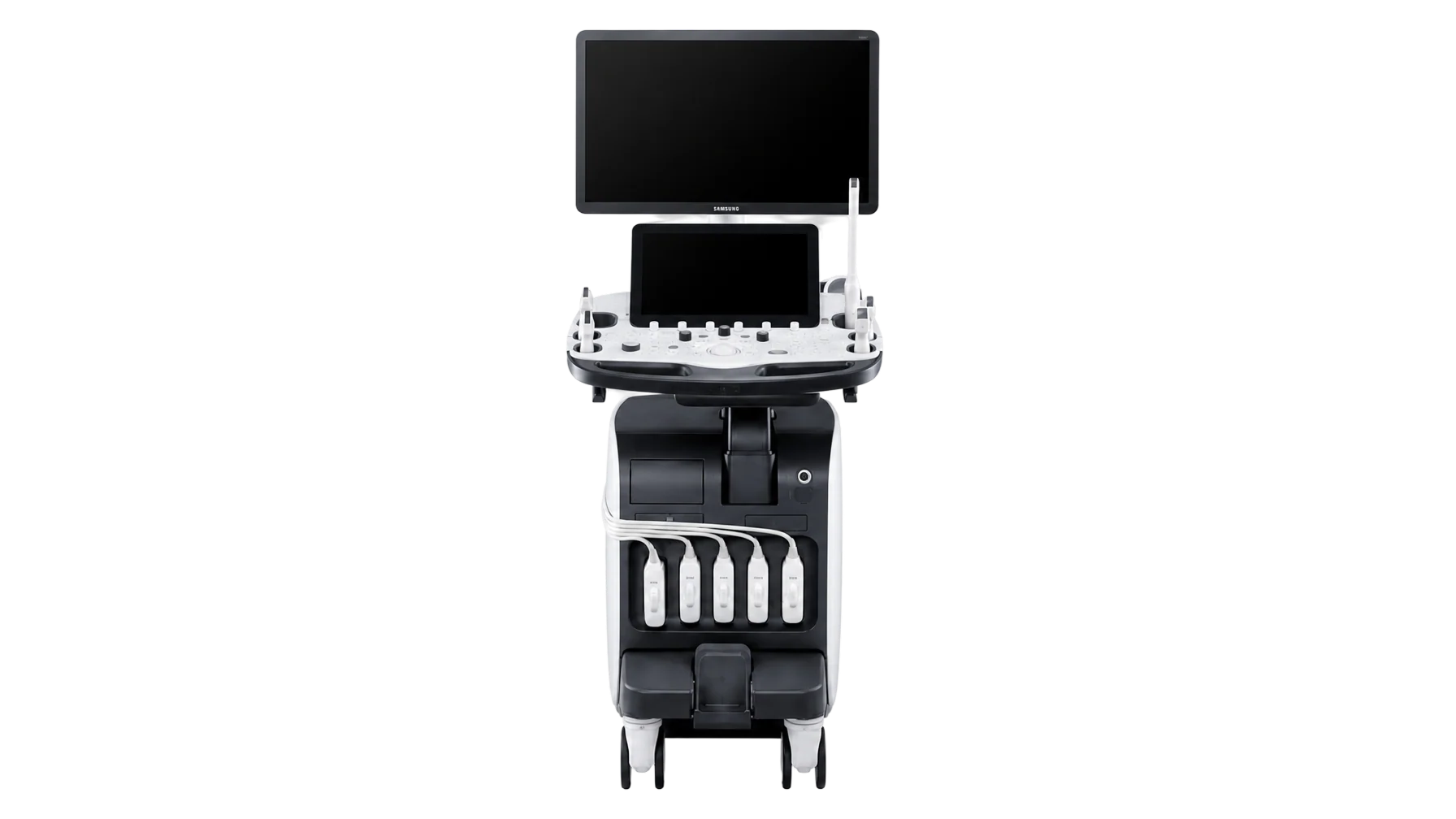
  • Carotid USCarotid UltrasoundCarotid wall thickness and plaque are examined to assess stroke risk.
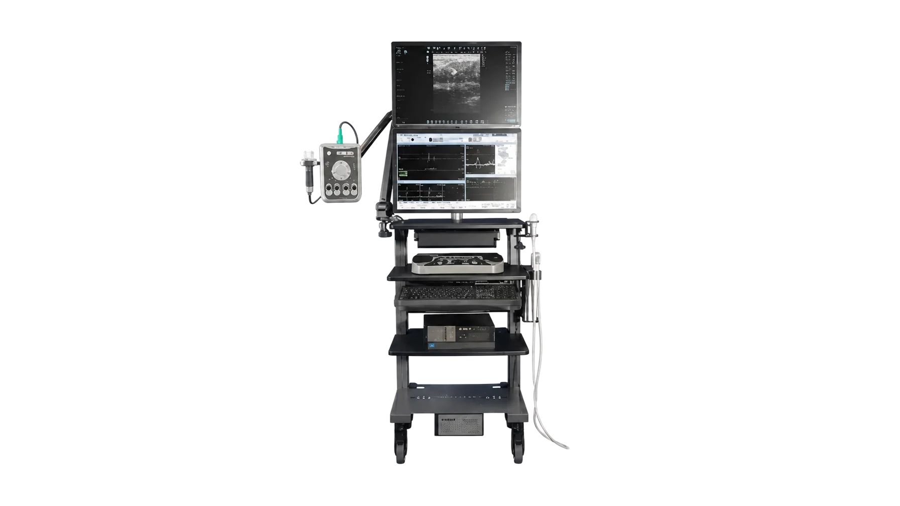
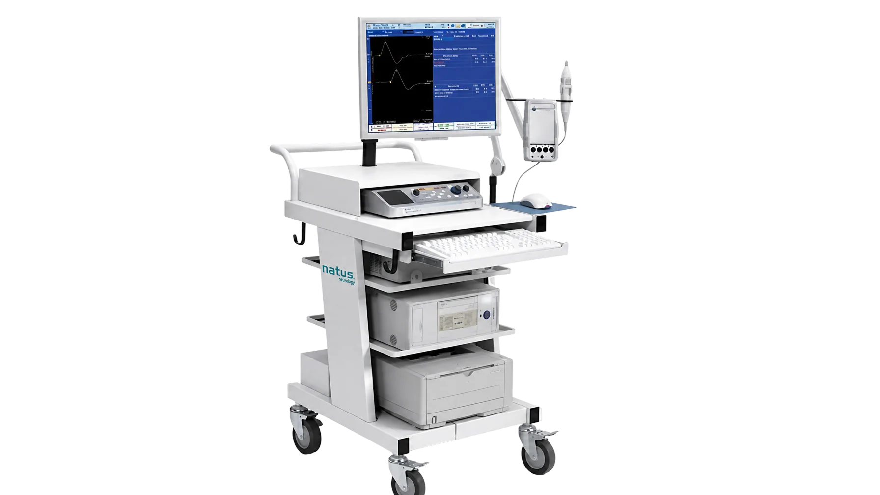
  • NCS / EMGNerve conduction studiesNerves in the arms, legs, and face are safely stimulated electrically and the responses recorded, establishing whether peripheral nerve damage is present and how severe it is.
  • EPEvoked potential testBy safely stimulating the visual and auditory pathways electrically, damage to the cranial nerves can be identified.
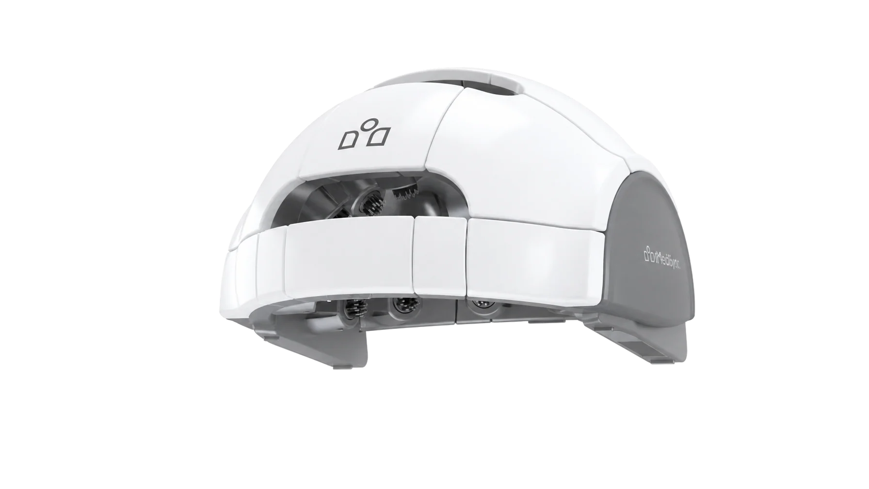
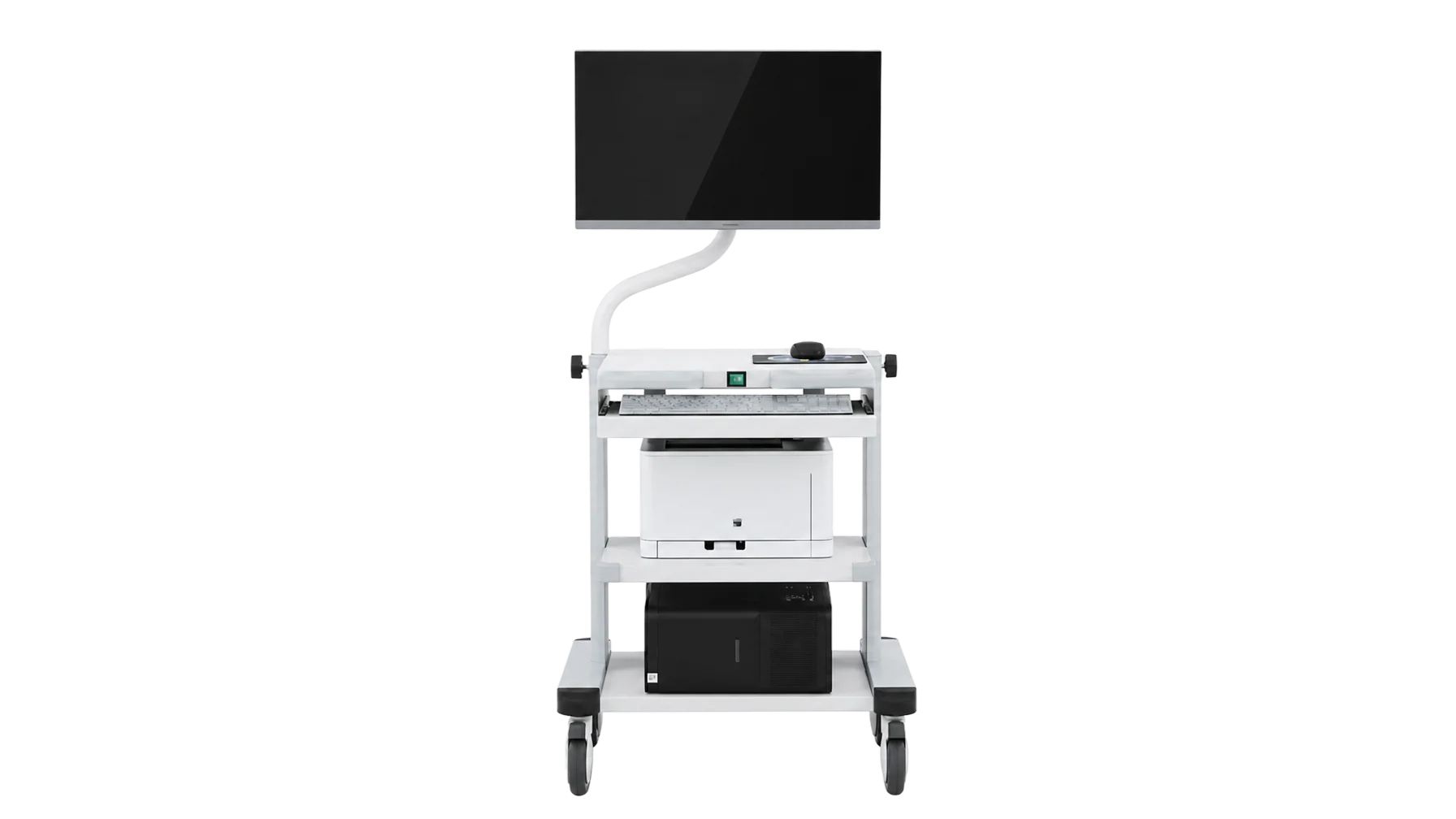
  • EEGAI digital EEGBeyond the conventional identification of epileptiform waveforms, quantitative EEG with the latest AI software aids in assessing brain function in headache, anxiety, and depression.
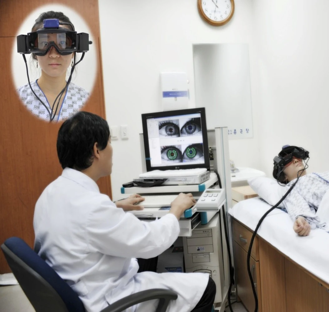
  • VNGVideo nystagmographyBy recording and analyzing involuntary eye movement on video, this test is useful for distinguishing dizziness disorders such as BPPV and Meniere's disease.
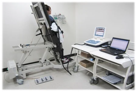
  • HRV / SSRAutonomic Function TestEquipment that objectively assesses the balance between the body's sympathetic and parasympathetic nerves.
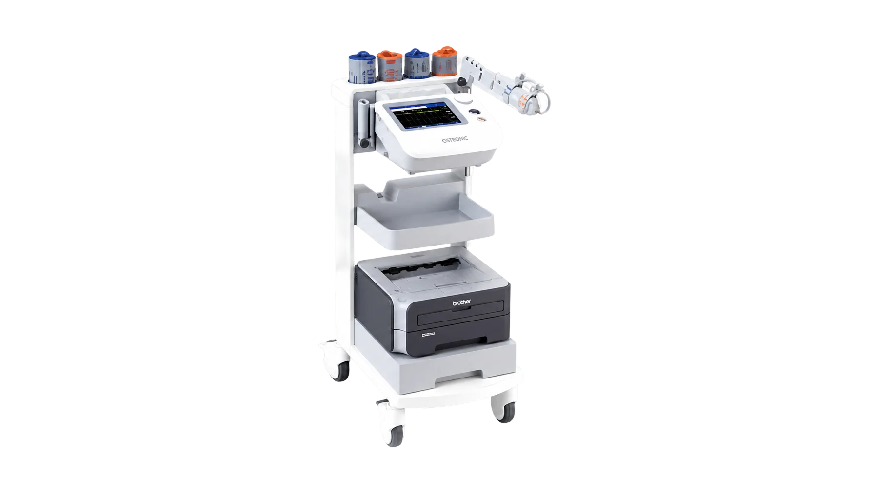
  • PWV / ABIArterial elasticity testingBy measuring vessel elasticity and the degree of atherosclerosis, it readily assesses the risk of circulatory disturbance and cardiovascular or cerebrovascular disease.
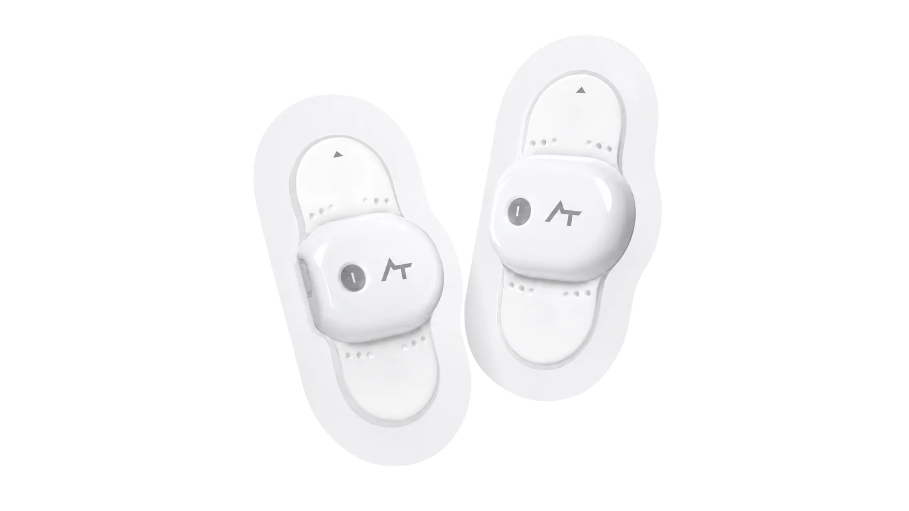
  • Wearable ECG/BPWearable cardiac function testingA micro-sensor continuously analyzes blood pressure and ECG over 24 hours to identify cardiac dysfunction capable of causing a stroke.
  • SNSB / CERADNeuropsychological testingIt distinguishes simple forgetfulness from dementia and objectively evaluates cognitive function including memory.
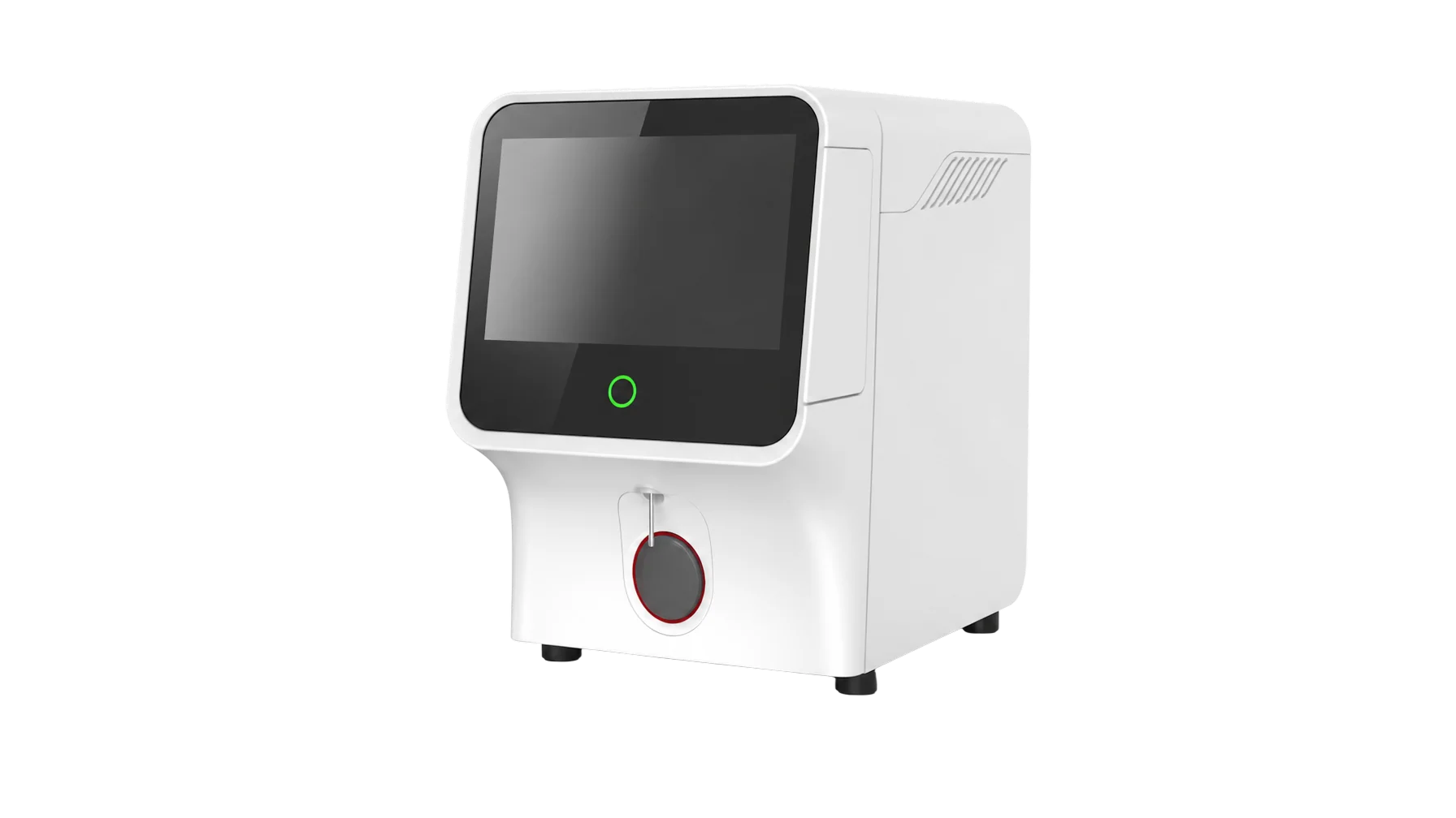
  • LABBlood and genetic testingWe identify risk factors for cardiovascular and cerebrovascular disease, including dyslipidemia, and look for high-risk dementia genes in advance so that prevention can begin.